

Over the past decade, foot and ankle surgery has undergone a quiet revolution. Procedures that once required long incisions and extensive soft-tissue dissection can now be performed through openings just a few millimeters wide. This approach — minimally invasive surgery, or MIS — has changed what recovery looks like for many of my patients. And now, a second transformation is underway: the arrival of artificial intelligence (AI) in the operating room.
Here is where the two are meeting, and what it means for the patients I treat.
Why minimally invasive surgery matters

For conditions like bunions (hallux valgus), the appeal of a minimally invasive approach is easy to understand. Compared with traditional open surgery, MIS offers:
- Faster recovery and a quicker return to daily life
- Smaller scars and a better cosmetic result
- Less soft-tissue disruption, which the body tolerates more gently
- Higher patient satisfaction in reported outcomes
These advantages are real, and they are the reason MIS has been adopted so widely for foot and ankle conditions.
The trade-offs surgeons work around
No technique is without limitations, and honesty about them is part of good care. Working through a few millimeters of skin means the surgeon operates with limited direct visualization — the small nerves and tendons just beneath the surface cannot be seen the way they can in open surgery. It also demands a high degree of precision performed largely by feel and by repeated X-ray (fluoroscopy) checks, and it carries a steep learning curve that rewards experience.
These are exactly the gaps that artificial intelligence is beginning to fill.
Where AI stands today
The research is growing quickly. AI-related publications in foot and ankle surgery have risen sharply — from a small fraction of the literature a decade ago to roughly a third of relevant papers in the most recent year. Today, AI is already contributing in several areas:
- Diagnosis and classification — assisting in the reading of ankle fractures and other injuries on imaging.
- Accurate, automated measurement — calculating bunion angles (such as the hallux valgus and intermetatarsal angles) consistently and objectively, including from weight-bearing CT.
- Surgical planning — powering 3D reconstruction of a patient’s anatomy for more tailored planning.
- Patient-specific implants — using load-bearing data to help design implants suited to the individual.
- Predicting outcomes — estimating recovery after fractures, sports injuries, and nerve injuries.
In short, AI is becoming a reliable second set of eyes for measurement and interpretation — the kind of objective, repeatable analysis that supports better surgical decisions.
Where the field is heading
The most exciting developments address the very limitations of MIS described above.
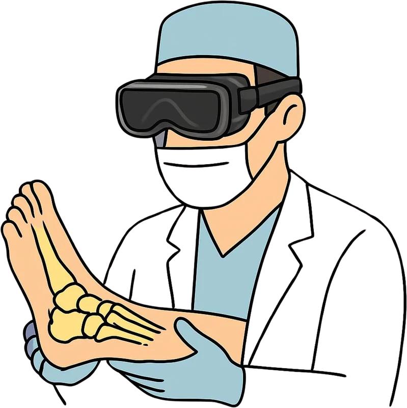
Seeing what can’t be seen. Cadaveric studies mapping the “danger zones” around key nerves and tendons are helping define safe corridors for instruments. Building on this, augmented and mixed reality overlays can register a patient’s pre-operative CT or MRI onto the live surgical field — effectively restoring the anatomic awareness that small incisions take away, and highlighting neurovascular structures the surgeon needs to protect.
Robotic navigation. Early clinical work is combining arthroscopy with robot-assisted, 3D-navigated screw placement. In one series of patients with talar neck fractures, this approach produced satisfactory outcomes. The concept is elegant: an intra-operative 3D scan aligns the imaging with the actual foot so they share a single coordinate frame; the surgeon plans the screw path on the scan, and the robotic arm precisely aligns the guide to that path.
Greater precision. AI-assisted image analysis also promises to reduce over- or under-correction during procedures like bunion surgery, moving beyond a surgeon’s subjective judgment toward objective, standardized targets.
The challenges that remain
I want to be candid: this technology is promising, not finished. Several real challenges must be solved before AI becomes routine:
- Limited data. Foot and ankle anatomy varies widely, and datasets remain relatively small.
- Trust and accountability. We need AI whose reasoning can be explained, and clear frameworks for who is responsible for AI-assisted decisions.
- Practical integration. Any tool must save time in the operating room, not add friction.
- Equity. Advanced systems are expensive, and we must guard against widening gaps in access to care.
What this means for you
For patients, the takeaway is reassuring. The goal of every one of these advances is the same: safer surgery, more precise correction, and faster recovery — with less guesswork and more objective, data-supported decision-making. Artificial intelligence will not replace the surgeon’s hands or judgment. It is becoming a powerful tool that makes an experienced surgeon safer and more precise.
Leave a Reply